Oocyte aging refers to the natural process by which a woman’s eggs (oocytes) age over time. This is no surprise because as we get older, so do all the cells that are born with us. Oocyte aging is the main cause of infertility for women in older age brackets.
As oocytes age, they face cellular changes that make it more difficult to conceive. These cellular changes can include:
1. DNA Damage: Over time, oocytes accumulate DNA damage, which can lead to an increased risk of chromosomal abnormalities in embryos.
2. Mitochondrial Dysfunction: Mitochondria are the energy-producing organelles in cells. Oocyte aging can result in mitochondrial dysfunction, which can affect the energy supply needed for fertilization and early embryo development.
3. Decline in Quantity and Quality: As women age, the number of viable oocytes decreases, and the remaining ones may have structural abnormalities or impaired function, reducing the chances of successful fertilization and healthy embryo development.
4. Telomere Shortening: Telomeres are protective caps at the ends of chromosomes. Oocyte aging is associated with telomere shortening, which can impact the ability of oocytes to divide properly during fertilization.
5. Cumulative Effects: These cellular changes can accumulate over time, making it more challenging for older women to conceive naturally and increasing the likelihood of pregnancy complications.
In summary, oocyte aging at the cellular level involves a range of biological changes that affect the quality, quantity, and functionality of a woman’s eggs, contributing to age-related declines in fertility and an increased risk of pregnancy-related issues. What if we can slow down this process, and maybe reverse it a little? It is no surprise that many women in advanced age brackets suffer from infertility and turn to donor eggs as an alternative option.
At North Cyprus IVF Center, we have been focusing on pregnancy in more advanced age brackets by offering treatments such as ovarian PRP, Mitochondrial Replacement Therapy, tandem IVF cycle and IVF with donor eggs. However, we are now introducing a break-through protocol that can combat age associated decline in female fertility and improve both oocyte count and quality in women in more advanced age brackets. For many years, supplementation with DHEA, myoinositol or antioxidant support have been the mainstay for treating women with diminished ovarian function. In recent years, we have seen some benefits with the use of human growth hormone, but this has also been quite limited in its reach.
Recent advances in anti-aging research provide us with deepening knowledge about how our cells function and more specifically, why they dysfunction. At the core of cellular aging lies epigenetic changes and cellular damage over time. Cellular aging can be slowed down or even reversed to some degree with interventions that can undo the damage acquired over time. Anti-aging research currently revolves around the role of sirtuins, NAD+ and senolytics when it comes to promoting cellular health and longevity.
Sirtuins are a group of proteins (specifically, SIRT1 to SIRT7) that play a crucial role in cellular health and longevity, and they are often associated with anti-aging processes. Here’s how they function in the context of antiaging:
1. DNA Repair: Sirtuins are involved in repairing damaged DNA, which is a key factor in aging. They help to maintain the integrity of the genetic code and prevent mutations that can lead to cellular dysfunction and aging.
2. Cellular Defense: Sirtuins promote cellular defense mechanisms, such as autophagy, which is the process of removing damaged or dysfunctional cellular components. This helps maintain cellular health and functionality.
3. Gene Regulation: Sirtuins influence gene expression by modifying histones, which are proteins that package DNA. By deacetylating histones, sirtuins can switch genes on or off, regulating various processes like metabolism, inflammation, and stress response.
4. Longevity Pathways: Sirtuins are linked to pathways like the NAD+ (nicotinamide adenine dinucleotide) system, which is crucial for energy metabolism and cellular function. Activating sirtuins with compounds like resveratrol or calorie restriction can potentially extend lifespan.
5. Cellular Stress Response: Sirtuins are involved in the body’s response to various stresses, such as oxidative stress and DNA damage. By enhancing the cellular stress response, they can help protect cells from age-related damage.
In summary, sirtuins are important regulators of various cellular processes that can impact aging. Activating sirtuins through lifestyle choices or potential therapies is a promising avenue for anti-aging research, as they can help maintain cellular health and potentially extend lifespan. Animal research shows that activating sirtuins can extend the lifespan by almost 30%. However, it’s important to note that while sirtuins show substantial benefits in animal studies, their role in human aging is still an area of ongoing research.
Both animal and human clinical studies show evidence for improved oocyte count and quality in IVF treatments1,2,3 after sirtuin pathways are activated.
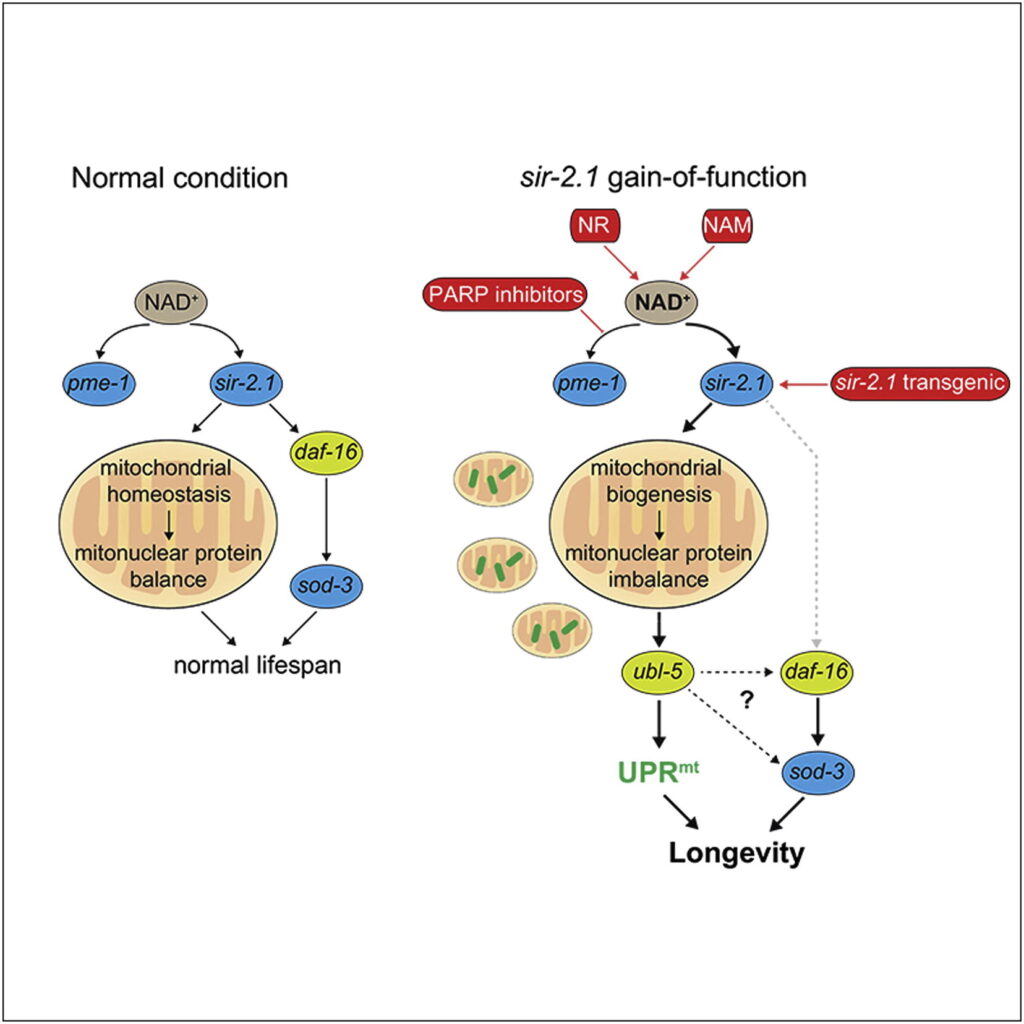
Nicotinamide adenine dinucleotide (NAD+) is a coenzyme that plays a vital role in various cellular processes. NAD+ is a critical molecule in cellular processes related to energy production, DNA repair, and cell signaling. Its levels and proper regulation are essential for maintaining cellular health, and researchers are exploring ways to modulate NAD+ levels as a potential strategy for promoting longevity and combating age-related diseases.
Recent animal and human clinical studies show significant positive correlation with increased NAD+ levels (through supplementation) and improved fertility as a result of improved oocyte parameters such as count, quality and fertilization capacity4,5.
Scientific research and clinical work show a clear benefit in IVF outcomes, providing support for the claims of cellular regeneration and improved longevity that have been attributed to the activation of sirtuin-NAD+ pathways.

Building upon scientific evidence, we have designed a specific protocol for oocyte anti-aging at North Cyprus IVF Center. This protocol involves activation of sirtuins via certain lifestyle changes such as intermittent fasting as well as sirtuin activating supplements for the duration of relevant parts of oogenesis. Similarly, our protocol involves increasing cellular NAD+ levels via oral supplementation as well as IV drips prior to the IVF procedure.
This protocol is mostly suited for women in more advanced age brackets (37+) who have diminished ovarian activity or a history of failed IVF cycles due to oocyte quality. Each patient’s protocol can slightly vary depending on their ovarian assessment. Therefore, the specific supplementation protocol is provided once our physicians assess your reproductive function. As a general idea, your protocol would involve some or all of the following:
1. Vitamin D
Vitamin D, a fat-soluble vitamin primarily synthesized in the skin upon sunlight exposure, plays an essential role in calcium homeostasis and immune function. Recent research has uncovered its importance in reproductive health, particularly regarding oogenesis, oocyte quality, and pregnancy outcomes. Vitamin D receptors (VDR) are expressed in human ovaries, suggesting a direct role in ovarian function (Irani & Merhi, 2014). Vitamin D appears to influence the maturation of follicles by regulating anti-Müllerian hormone (AMH) levels, a marker of ovarian reserve (Paffoni et al., 2014). Moreover, studies have indicated that higher vitamin D levels are associated with better oocyte quality and fertilization rates in women undergoing IVF (Rudick et al., 2012). Vitamin D’s role in oocyte quality is partly due to its anti-inflammatory and antioxidant properties, which protect oocytes from oxidative stress. Several studies have demonstrated that adequate vitamin D levels are also linked to improved pregnancy outcomes. For example, higher serum vitamin D concentrations have been associated with higher clinical pregnancy and live birth rates in women undergoing assisted reproductive technology (ART) (Rudick et al., 2012). Additionally, vitamin D deficiency has been linked to pregnancy complications such as preeclampsia, gestational diabetes, and preterm birth (Bodnar et al., 2007).
2. Nicotinamide Mononucleotide (NMN)
Nicotinamide Mononucleotide (NMN) is a precursor to NAD+ (Nicotinamide Adenine Dinucleotide), a coenzyme involved in cellular energy metabolism and DNA repair. NAD+ levels naturally decline with age, which can impair oocyte quality and fertility (Imai & Guarente, 2014). NMN has been shown to improve mitochondrial function and energy production within oocytes, which is crucial for their maturation and development (Bertoldo et al., 2020). Studies in animal models have demonstrated that NMN supplementation can rejuvenate aged oocytes by restoring mitochondrial activity and increasing NAD+ levels, leading to improved oocyte quality (Bertoldo et al., 2020). This is particularly relevant in older women, where mitochondrial dysfunction contributes to declining fertility. Although human clinical trials are still limited, animal studies suggest that NMN supplementation can enhance fertility and pregnancy outcomes. By improving mitochondrial function and reducing oxidative damage, NMN helps preserve oocyte quality, leading to better embryo development and increased pregnancy rates (Bertoldo et al., 2020). These findings are promising for women experiencing age-related fertility declines.
3. Fisetin
Fisetin is a naturally occurring flavonoid found in various fruits and vegetables. It has potent antioxidant, anti-inflammatory, and senolytic properties, meaning it helps clear out senescent cells, which accumulate with age and contribute to reproductive aging (Yousefzadeh et al., 2018).
Fisetin’s antioxidant properties help protect oocytes from oxidative stress, a major contributor to oocyte aging and quality decline. Oxidative stress can damage the DNA and mitochondria within oocytes, leading to chromosomal abnormalities (Liu et al., 2020). By reducing oxidative damage and clearing senescent cells, fisetin can support healthier oocyte maturation and enhance oocyte quality (Yousefzadeh et al., 2018). Although research on fisetin’s direct effects on pregnancy outcomes is still in its early stages, its ability to reduce inflammation and oxidative stress may improve overall reproductive health. Studies suggest that reducing senescent cells in the ovarian environment may lead to better follicular development, which could positively impact pregnancy outcomes (Liu et al., 2020).
4. Glycine and N-Acetylcysteine (GlyNAC)
Glycine is a conditionally essential amino acid with roles in collagen synthesis, antioxidation, and metabolic regulation. N-Acetylcysteine (NAC) is a precursor to glutathione, one of the body’s most powerful antioxidants, which plays a critical role in protecting cells from oxidative stress.
Glycine and NAC can work synergistically to enhance oocyte quality by mitigating oxidative stress and supporting mitochondrial health. NAC helps increase intracellular glutathione levels, which directly protect oocytes from oxidative damage (Elizur et al., 2009). Oxidative stress is particularly detrimental during oogenesis, leading to poorer oocyte quality and increased chromosomal abnormalities (Guerin et al., 2001). Glycine, on the other hand, helps stabilize the mitochondrial membrane potential and may assist in maintaining intracellular calcium levels, which are critical for oocyte maturation and fertilization (Yoon et al., 2014). NAC has been studied in women with polycystic ovary syndrome (PCOS), where it has been shown to improve ovulation rates and pregnancy outcomes by reducing insulin resistance and oxidative stress (Fulghesu et al., 2002). Additionally, glycine’s role in cellular protection and its antioxidative properties may contribute to improved embryo quality and increased implantation rates in ART (Yoon et al., 2014).
5. Alpha-Lipoic Acid (ALA)
Alpha-Lipoic Acid (ALA) is a potent antioxidant involved in mitochondrial energy metabolism and the reduction of oxidative stress. It is both water- and fat-soluble, allowing it to act within both the cellular membrane and the cytoplasm (Packer et al., 1995).
ALA has been shown to improve mitochondrial function, which is crucial for oocyte quality and energy production during oogenesis. It acts as a cofactor for mitochondrial enzymes, helping to generate ATP, the energy required for oocyte maturation (Güney et al., 2015). By reducing oxidative stress, ALA also helps preserve the integrity of oocyte DNA and cellular structures (Jiang et al., 2020). ALA’s ability to improve mitochondrial function and reduce oxidative damage may enhance embryo quality and increase pregnancy success rates in ART. Studies suggest that ALA supplementation can improve ovarian response in women with diminished ovarian reserve, leading to better oocyte quality and improved chances of conception (Güney et al., 2015).
Supplements need to come from companies that are GMP-compliant, and third party tested. Therefore, we only recommend the following brands:
1- Do-Not-Age: This brand comes up at number one on the supplement companies list due to purity of its products. If purchasing from this company, don’t forget to use ELITE10 as your coupon code for a 10% discount.
2- VitalityPRO: This brand is also third-party tested and transparent about its products purity. If purchasing from this company, don’t forget to use ELITE10 as your coupon code for a 10% discount.
3- Nature’s Fusions: If purchasing your products from the United States, Nature’s fusions is one of the best suppliers which guarantees purity. You will need to contact them for a discount code before placing a purchase by mentioning you have been referred by Dr. Ahmet Ozyigit at Elite Hospital.
References
1. Okamoto, N. et al. (2022) ‘Short-term resveratrol treatment restored the quality of oocytes in aging mice’, Aging, 14(14), pp. 5628–5640. doi:10.18632/aging.204157.
2. Vo, K.C., Sato, Y. and Kawamura, K. (2023) ‘Improvement of oocyte quality through the sirt signaling pathway’, Reproductive Medicine and Biology, 22(1). doi:10.1002/rmb2.12510.
3. Iljas, J.D., Wei, Z. and Homer, H.A. (2020) ‘SIRT1 sustains female fertility by slowing age‐related decline in oocyte quality required for post‐fertilization embryo development’, Aging Cell, 19(9). doi:10.1111/acel.13204.
4. Pollard, C.-L. et al. (2022) ‘NAD+, sirtuins and parps: Enhancing oocyte developmental competence’, Journal of Reproduction and Development, 68(6), pp. 345–354. doi:10.1262/jrd.2022-052.
5. Bertoldo, M.J. et al. (2020) ‘NAD+ repletion rescues female fertility during reproductive aging’, Cell Reports, 30(6). doi:10.1016/j.celrep.2020.01.058.
6. Bodnar, L. M., Catov, J. M., Zmuda, J. M., Cooper, M. E., Parrott, M. S., Roberts, J. M., &
7. Marazita, M. L. (2007). Maternal serum vitamin D levels and pregnancy outcomes. American Journal of Obstetrics and Gynecology*, 197(3), 1-7.
8. Elizur, S. E., Lebovich, M., Sellman, L., Agarwal, A., & Ashok, A. (2009). N-acetylcysteine supplementation in PCOS. *Reproductive BioMedicine*, 18(5), 61-70.
9. Fulghesu, A. M., Ciampelli, M., Muzj, G., Belosi, C., Selvaggi, L., Lanzone, A., & Apa, R. (2002). N-acetylcysteine treatment improves insulin sensitivity. *Fertility and Sterility*, 77(6), 1128-1135.
10. Güney, M., Oral, B., Karahan, N., Mungan, T., & Kapucuoglu, N. (2015). Alpha-lipoic acid improves oocyte quality and mitochondrial function. *Fertility and Sterility*, 91(6), 2563-2569.
11. Irani, M., & Merhi, Z. (2014). Vitamin D and oogenesis: A potential role in ovarian function. *Reproductive Biology and Endocrinology*, 12, 86.
12. Jiang, H., Tian, S., & Zheng, S. (2020). Antioxidant effects of alpha-lipoic acid on mitochondrial health in reproduction. *Mitochondrion*, 51, 1-8.
13. Liu, B., Zhou, Z., Zhou, W., Lin, J., & Zhou, Y. (2020). Effects of fisetin on oocyte aging and mitochondrial function. *Journal of Cellular Physiology*, 235(1), 153-162.
14. Packer, L., Witt, E. H., & Tritschler, H. J. (1995). Alpha-lipoic acid as a biological antioxidant. *Free Radical Biology and Medicine*, 19(2), 227-250.
15. Paffoni, A., Ferrari, S., Viganò, P., Pagliardini, L., Papaleo, E., Candiani, M., & Fedele, L. (2014). Vitamin D status and ovarian reserve in


